FLAME member achieves enormous progress in electron microscopy
2019/04/08 by Christian Meier
Darmstadt researchers led by Leopoldo Molina-Luna observe the tiniest of electronic components in action. Not only can their electron microscope image individual atoms; it can also produce, heat and electronically control the sample.
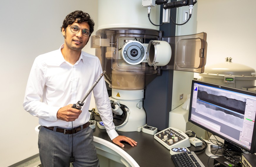
Although the two control boxes at the bottom of the room-sized electron microscope are hardly noticeable amongst the numerous screens and cables, they make all the difference for Leopoldo Molina-Luna. They serve as an upgrade that turns the microscope into an entire laboratory for nanotechnology. The physicist wants more than just to observe the world of individual atoms. He wants to manipulate it. ‘The goal is to understand what actually happens on the nanoscale,’ says Molina-Luna, expert in electron microscopy at the Department of Materials and Earth Sciences. When they use the term ‘nano-level’, scientists are referring to an order of magnitude in which objects measure only a few nanometres – no more than one hundred thousandth of a millimetre. In this minuscule universe, an influenza virus is already a giant. However, technology has been able to contract into this world for some time. The smallest elements of a computer chip, for example transistors, measure only a few nanometres. Billions of them fit on a fingernail-sized chip.
Nanoelectronics wants to break new ground, such as components with a similar reminder function as brain cells or storage media with gigantic capacity in the tiniest space. For this, different materials must be added to layers that are only a few nanometres thin: custom work in the invisible microcosm.
A test lab, just a few millimetres in size
Although you can see such diminutive components in the electron microscope, you only glimpse snapshots. However, Molina-Luna wants to watch the tiny electronic components while they are at work. He wants to observe how individual atoms behave during the switching process. Such a component consists of a thin insulator slice between two metal layers, not unlike a sandwich. When electrical voltage is applied, a conductive bridge forms between the metals, a so-called filament, which can be interrupted again by changing the applied voltage. These two different states allow the storage of data if one encodes one state as 0 and the other as 1.
‘At least, that is the model concept,’ says Molina-Luna. ‘Until now, it could only be tested indirectly, because classical measurement methods are macroscopic.’ Current methods measure the current flow at a certain voltage, a phenomenon collectively caused by tens of billions of particles. ‘But electron microscopy has made tremendous progress in recent years,’ says Molina-Luna. The electron microscope has become a ‘multidimensional’ instrument.
The development of so-called Micro-Electro-Mechanical Systems (MEMS) helped. These are complex electromechanical devices of microscopic size. Molina-Luna‘s cooperation partner, the Dutch company DENSsolutions, has shrunk a test lab down to a few millimetres and fixed it on a sample holder. This chip can be inserted into an electron microscope. This now allows the Darmstadt researchers to heat samples within the microscope and apply different electrical voltages. The sample holder is very stable, emphasizes Molina-Luna. It allows very high magnifications, up to 25-million times. At the same time, the sample holder serves as a production site: The Darmstadt researchers have synthesized nanoparticles on it. The powder was mixed with the solvent, the solvent could evaporate, and a sintering process took place after introducing the holder in the microscope, which produced the ‘new’ nanoparticles.
Mechanism of flexoelectricity in nanoparticle elucidated
Recently, researchers have observed an effect on such particles, which could facilitate new, super-dense storage media. With a core and a shell, the nanoparticles resemble cherries. The core consists of a different metal oxide than the shell (the core of sodium bismuth titanate, the shell of strontium-rich sodium bismuth titanate). When it is forced into a particle, mechanical stress builds up in its interior. Researchers controlled the strength of the stress by changing the proportion of strontium.
The mechanical stress leads to so-called flexoelectricity: Inside, the electrical charge carriers split into positive and negative charge centres, creating an electric dipole. Through flexoelectricity, this is retained even at high temperatures, something the Darmstadt researchers recently observed in this system for the first time anywhere in the world. Thanks to their microscope, they were able to elucidate the mechanism of flexoelectricity in the nanoparticle. The effect is shown in tiny patterns resembling fishbones. Researchers published the results in the renowned journal Nature Communications.
Because flexoelectricity is minimal in traditional materials, it has received little attention so far. However, it could be technically interesting, specifically if it could be switched on and off, because switching between two states could encode a bit and thus act as a memory. The TU researchers showed that this is possible: The polarization in their nanoparticles could be switched by applying electrical voltage, also inside the microscope. In addition to being used as memories, flexoelectric components could serve as sensors, actuators or energy converters.
With their success, the Molina-Luna team demonstrated how flexible the new microscopy method is. ‘ Such experiments help us to develop the chip even further,’ says Molina-Luna. The extended form of microscopy will be applied to several classes of nanomaterials. Molina-Luna has received one of the prestigious ERC Starting Grants from the European Research Council for the project. The Darmstadt laboratory has the technology needed to prepare samples. With a beam of accelerated ions, Molina-Luna‘s team can cut very thin layers from components so that they can be examined using the electron microscope. Though the Darmstadt scientists see themselves as basic researchers, they are still looking for materials ‘suitable for the application,’ says Molina-Luna.
Molina-Luna regards extended electron microscopy as a complement to previous methods, such as the testing of samples with x-rays or electrical measurement. ‘We contribute to the overall picture,’ he says. Information on what is happening at the atomic level is frequently still lacking today. For example, regarding filament-based memories, one can now directly see the role that oxygen atoms play in forming a filament through an insulating layer. ‘What is new is that we can now say how the switching process happens,’ says the physicist. ‘We want to refine the models,’ he adds. But this does not complete the claims of the researchers: ‘Ultimately, it‘s about developing better devices.’
At a glance
EU-Förderung: European Research Council (ERC) Starting Grant: Functionality of Oxide based devices under Electricfield: Towards Atomicresolution Operando Nanoscopy (FOXON), Budget ca. 1,8 Millionen Euro.
Publication: Enabling nanoscale flexoelectricity at extreme temperature by tuning cation diffusion.Molina-Luna, Leopoldo et. al. In: Nature Communications