Crystal Structure, Structure Research Division
Head: Prof. Dr. Wolfgang Donner
The main focus of structural research is the characterization of powders and single crystals by X-ray diffraction. With this method, modulations and phase transitions can be investigated and the electron density can be calculated by the determination of structure factors. The fabrication of single crystals of different sizes allows us and other subproject groups to characterize defect free samples, providing accurate information on atomic and electronic structure. The aim is to develop a model that combines the structure with the properties and provides a better understanding of the antiferroelectric phase.
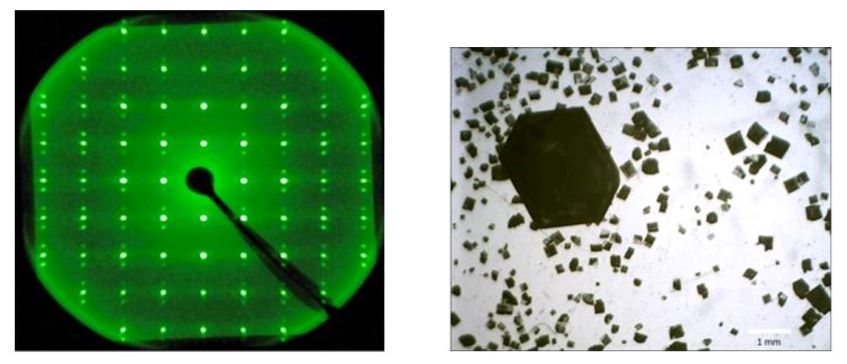

[1] J Dec Real domain structure in orthorhombic phase of NaNbO3 crystals, Crystal Res. & Technol. 18, 195 (1983)
Local Structure, Physical Chemistry Division
Head: Prof. Dr. Gerd Buntkowsky
We are interested in the relation between the local structure and the functional properties of lead-based and lead-free antiferroelectric (AFE) perovskites. Solid-state nuclear magnetic resonance spectroscopy (NMR) is used to investigate the structural changes occurring through the AFE-FE phase transition as a function of temperature and chemical composition, since this phase transition is closely associated with the functional properties. From the analysis of NMR observables (chemical shift, electric field gradient), we aim at a deeper understanding of these structure-property relations. This knowledge will complement structural information obtained by spectroscopic techniques and diffraction experiments.
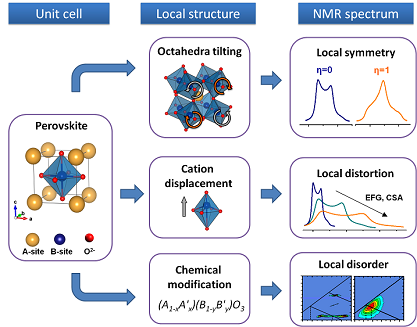
Furthermore, local structure parameters determined by NMR will allow the development of structural models and serve as a reference for the evaluation of results obtained from theoretical calculations. The obtained knowledge of the relation between local structure and macroscopic properties in lead-based AFEs will further assist in the rational design of lead-free alternative materials.
Transmission Electron Microscopy, Advanced Electron Microscopy Division / Geomaterial Science Division
Head: Prof. Dr. Leopoldo Molina-Luna / Prof. Dr. Hans-Joachim Kleebe
Transmission electron microscopy (TEM) is a powerful technique to investigate the microstructure of solid ceramics and thin film samples and to establish structure-property correlations on the atomic scale. By using electron diffraction based methods, as well as conventional and aberration (Cs) corrected (S)TEM imaging, an insight into the domain structure and local crystallographic orientations can be obtained. In conjunction with spectroscopic methods (EDX, EELS), scanning transmission electron microscopy (STEM) also allows the determination of chemical inhomogeneities with high spatial resolution at grain and phase boundaries. In addition, we plan to use a state-of-the-art in situ TEM sample holder for temperature dependent studies at high-spatial resolution. Furthermore, by applying an electric-field directly inside the microscope, domain formation studies during the AFE → FE phase transition will be possible.
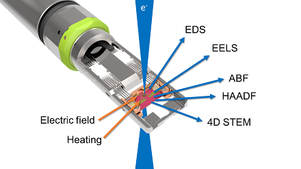
In conjunction with spectroscopic methods (EDX, EELS), scanning transmission electron microscopy (STEM) also allows the determination of chemical inhomogeneities with high spatial resolution at grain and phase boundaries. In addition, we plan to use a state-of-the-art in situ TEM sample holder for temperature dependent studies at high-spatial resolution. Furthermore, by applying an electric-field directly inside the microscope, domain formation studies during the AFE → FE phase transition will be possible.